This is the fifth in a series on the senses. Past articles in this series included: an introduction to the variety of senses, Chemoception, Thermoception and Mechanoception. This article is something of an anomaly given the theme. You see, I was planning to write about the sense of vision – the ability to sense a very narrow band of photons with energies of around 3 to 1.5 electron volts (eV) and wavelengths between 400–700 nanometers – and call it photoreception. But logic raised an eyebrow and questioned the term “photoreception” in the title. Recall high school physics lectures on photons, and you will remember that photons are electromagnetic moieties where lower wavelengths like gamma rays have tiny wavelengths and higher energies (0.01 nm and 100 keV), grading to radio waves with enormous wavelengths and tiny energies (>1000 km and 10-15 eV) (see Figure 1). The technical definition of photons is that they are force-carrying bosons which transmit energy between atoms in our universe. Einstein famously showed that they can exist as both a particle and a wave. It was for demonstration of the photoelectric effect – the ability of a photon carrying a specific quantum of energy to induce a current in a metal plate – that Einstein received his one and only Nobel Prize. [1]
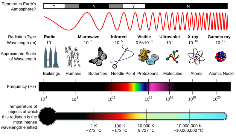
Figure 1: A diagram of the electromagnetic spectrum, showing various properties across the range of frequencies and wavelengths. Attributed to: By Inductiveload, NASA – self-made, information by NASA. CC BY-SA 3.0, https://commons.wikimedia.org/w/index.php?curid=2974242
It is obvious from Figure 1 that the term “photoreception” should equally apply to the ability to sense photons of gamma radiation, ultraviolet, infrared (heat), microwaves and radiowaves. Although no known life-form can sense the very low and very high wavelengths, arthropods and many vertebrates (although not humans) can see in ultraviolet [2] and pit vipers [3] see in infra-red. Clear fossil evidence of eyes dates back to 530 Ma to the Cambrian explosion, when the variety of animal phyla that exist today emerged, including arthropods and vertebrates. [4] When they started seeing and hunting each other, some developed tough armor to resist attack and others sharp cutting tools to break through the armor. These hard bodies fossilized, which is how we know they existed and had eyes. But animals existed long before that; they were soft-bodied jellyfish and worms, and did not fossilize so well. Today’s box jellyfish have eyes, and other jellyfish have light-sensing patches, so the ability to sense light and detect things around them probably predates 530 Ma. Eyes also predated the brain, according to some hypotheses, since the brain is built around information processing and complex information was not sensed before vision became available. [5] In fact, box jellyfish have camera eyes of the type that vertebrates and cephalopods have, but they lack a brain.
So what is sight/vision in animals? It is the ability to activate neurons that project from the retina to the brain’s visual cortex, where an image is formed reflecting the light that fell on the retina. The earliest eye, according to Darwin, would have been as simple as a nerve coated with a photosensing pigment. All eyes share some genetic similarities, so they probably evolved from a single ancestor despite what seem like profound differences today. A schematic of the structural and functional evolution of eyes is shown in Figure 2.
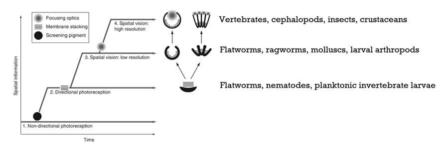
Figure 2: Sequential evolution of the four classes of sensory tasks controlled by light and corresponding innovations in eye design. Class 1 tasks require basic sensing of light and dark; Class 2 tasks require directional sensitivity for phototaxis and alarm responses; Class 3 tasks are self-motion, habitat selection and orientation to landmarks or celestial objects; Class 4 tasks are detection and pursuit of prey, predator evasion, mate detection and evaluation, visual communication and other sophisticated visual tasks. Note that increasing directionality and resolution are due to increased spatial vision, and clarity is due to lenses that focus light on photosensitive retinas. Modified from Reference [4] under fair use for educational purposes.
Now, imagine you and a bee looking at the same flower. The structures of your eyes are different, as shown in Figure 3. How light is processed in each is described in Table 1.

Figure 3: Details of a human’s eye and retinal structure and a bee’s eyes and compound eye structure. The 3 ocelli at the top of the head are used for sensing light polarization rather than for vision per se. The human retina image is reproduced from Reference [6], and the bee eye is modified from Reference [7], under fair use for educational purposes.
Table 1: How light is processed in human eyes vs. bee eyes.
| Human | Bee |
|---|---|
| Light enters the domed cornea, which bends the light inwards towards the pupil, which shrinks or dilates to adjust light intensity. Light passes through the crystalline lens, which bends it to focus on the photoreceptor cells. Photoreceptor cells are nerve cells which come in two forms: rods (with rhodopsin, which works in dim light) and cones (which work in bright light and colour vision). Cone opsins come in three varieties: S (blue detection), M (green detection) and L (red detection), and detect across the 400–700 nm range. Each opsin within a rod or cone cell is associated with 11-cis-retinal, the chromophore which absorbs light. When a photon of light hits the rod or cone, the straight 11-cis-retinal transforms to bent 11-trans-retinal. This causes the bent retinal molecule to push against the opsin and cause it to change shape. Opsins activate a signal cascade which amplifies the signal many thousands of times over. The action then moves out of the photoreceptor cell and passes through two layers of nerve cells called bipolar cells and ganglion cells, which process the signal by identifying motion, detecting contrast and edges of images. The axons of the ganglion cells bundle into the optic nerve, which sends action potentials to the visual cortex of the brain via the thalamus. The brain processes the image and fills in gaps to provide a smoothed-out image. | Two types of eyes: two compound eyes with ommatidia, and three simple ocelli at the top of the head for detecting light polarization. The compound eyes, which process images, are discussed here. The roughly 5,000 chambers called ommatidia of each eye each have a lens which points in a slightly different direction. Light from the flower enters all the ommatidia and focuses on a cluster of photoreceptor cells called the rhabdom. Each ommatidium therefore contributes just one point of light, or one ‘pixel’, to the mosaic image. Each ommatidium of the compound eye forms a long, narrow tube. At the top is a corneal lens (the facet in Figure 3), and below that is a crystalline cone which focuses the image. Each ommatidium contains typically eight elongated photoreceptor cells containing retinal-linked opsins, with tiny projections called microvilli along the inside which make contact to form the rhabdom, the light-sensing core. Bees also have three opsins, but these detect ultraviolet, blue and green (300–650 nm). They do not sense the red wavelength. When light hits the facet, it travels down through the corneal lens, gets focused by the crystalline cone, and then reaches the rhabdom. The light triggers the flat 11-cis-retinal to isomerize to bent 11-trans-retinal, which pushes the opsin and activates photoreceptor neurons that project into the optic lobe. The optic lobe processes the images and consists of the lamina (contrast and timing), the medulla (colour, motion and polarization) and the lobula (feature detection and motion processing), which refine the image. The image is then transmitted to the bee’s central brain for integration with other senses and for responding. |
The output [8]: University of Exeter scientists built a bee’s-eye view that distinguishes between flowers by combinations of colors and patterns. Bees focused more on the patterns than the colors, although they could distinguish both. [9] The patterns and colors as detected by bees vs. humans are shown in Figure 4. A video about the structural and functional aspects of bee eyesight is presented in Figure 5.
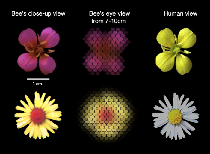
Figure 4: Flowers as seen by human eyes and bees’ eyes at various distances. Figure credit: Natalie Hempel de Ibarra (University of Exeter). Reproduced from Reference [8] under fair use for educational purposes.
Here is a short video of bee vision:
Figure 5: Video about the anatomical and functional aspects of bee eyesight.
References
[1] Einstein’s 1921 Nobel Prize citation read “for his services to Theoretical Physics, and especially for his discovery of the law of the photoelectric effect”. This was an egregious slight, given that Einstein’s “miracle year” of 1905 saw him publish not one but three paradigm-shifting papers: (1) on the existence and size of the atom (hypothesized by Democritus around 400 BCE); (2) Special Relativity, showing time and space were relative, that mass was just another manifestation of energy (famously including the formula E=mc2), and that the speed of light is a maximum that nothing can exceed; and (3) the photoelectric effect, mentioned above. In addition, in 1915 Einstein shook the foundations of science with his General Theory of Relativity, which postulated that gravity is a bending of space. The experimental demonstration by Eddington in 1919 was well before the 1921 Nobel Prize, and the omission of this from the citation was unconscionable.
[2] Cronin, T.W. and Bok, M.J. (2016). Photoreception and vision in the ultraviolet. Journal of Experimental Biology, 219(18), pp.2790–2801. doi:10.1242/jeb.128769.
[3] Newman, E.A. and Hartline, P.H. (1982). The infrared “vision” of snakes. Scientific American, 246(3), pp.116–127.
[4] Land, M.F. and Nilsson, D-E. (2026). Animal Eyes. Available at: https://books.google.com/books?id=vQgWDAAAQBAJ [Accessed 26 Apr. 2026].
[5] Gehring, W.J. (2005). New Perspectives on Eye Development and the Evolution of Eyes and Photoreceptors. Journal of Heredity, 96(3), pp.171–184. doi:10.1093/jhered/esi027.
[6] Social Sci LibreTexts (2025). 6.3: The Retina. Available at: https://socialsci.libretexts.org/Bookshelves/Psychology/Biological_Psychology/Behavioral_Neuroscience_(OpenStax)/06:_Vision/6.03:_The_Retina [Accessed 26 Apr. 2026].
[7] Museum of the Earth (n.d.). Bee Biology. Available at: https://www.museumoftheearth.org/bees/biology.
[8] EurekAlert! (2022). Flowers as seen by human eyes and bees’ eyes at various distances. Available at: https://www.eurekalert.org/multimedia/948552.
[9] Hempel de Ibarra, N., Holtze, S., Bäucker, C., Sprau, P. and Vorobyev, M. (2022). The role of colour patterns for the recognition of flowers by bees. Philosophical Transactions of the Royal Society B, 377(1862). doi:10.1098/rstb.2021.0284.
